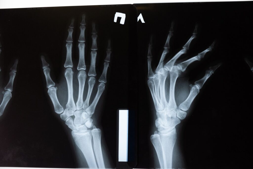
Dual-energy X-ray absorptiometry, usually shortened to DXA or DEXA, is one of the better established tests in the longevity-testing orbit. It measures bone mineral density, can estimate regional fat and lean mass, and increasingly appears in private “body age” packages. The clinical signal is real. The mistake is treating one scan as a verdict on ageing, fracture destiny, or metabolic health.
What DXA was built to measure
DXA’s strongest use remains bone. The scan sends two low-dose X-ray beams through the body and estimates how much mineral is present in a defined area of bone, most often the hip and lumbar spine. That result is reported as bone mineral density, then compared with a young-adult reference range to produce a T-score.
The reason clinicians care is not aesthetic. Lower bone mineral density is associated with higher fracture risk, and fractures in later life are not minor events. Hip fractures, vertebral fractures, and wrist fractures can change mobility, independence, and mortality risk. A DXA scan is therefore best understood as a risk-stratification tool, not a photograph of how “old” a skeleton is.
The latest US Preventive Services Task Force recommendation is clear on the best-supported screening group: women aged 65 or older should be screened for osteoporosis with DXA bone mineral density, with or without fracture-risk assessment. It also recommends screening postmenopausal women under 65 when risk factors and formal risk assessment suggest increased risk. For men, the task force found the evidence insufficient to judge population screening benefits and harms.
That distinction matters. DXA is not a universal annual health MOT. Its value rises when the person being scanned has a realistic pre-test probability of low bone density or high fracture risk.
The T-score is useful, but incomplete
The best-known DXA number is the T-score. A T-score of -2.5 or lower at the hip or spine is commonly used to define osteoporosis. A score between -1.0 and -2.5 is often labelled osteopenia, or low bone mass.
Those thresholds are useful, but they can make the test look more binary than it is. Bone density sits on a continuum. A person with a T-score of -2.4 is not biologically safe whilst a person at -2.5 is suddenly fragile. The result needs context: age, sex, previous fractures, steroid exposure, smoking, alcohol intake, rheumatoid arthritis, parental hip fracture, falls risk, and other conditions all change the interpretation.
This is why fracture-risk calculators exist. The FRAX tool was developed to estimate fracture risk by combining clinical risk factors with, where available, femoral-neck bone mineral density. In the UK, NICE advises clinicians to assess fragility-fracture risk using tools such as FRAX or QFracture, which estimate absolute risk of hip and major osteoporotic fracture. DXA can sharpen the estimate, but it does not replace the clinical picture.
The practical question is rarely “what is my score?” It is “does this score change what I should do?”
Where body-composition DXA helps
Whole-body DXA can also estimate fat mass, lean mass, bone mineral content, and regional distribution. This is why private clinics market it to athletes, people losing weight, and adults worried about visceral fat or sarcopenia.
Compared with bathroom scales and bioelectrical impedance devices, DXA is a more controlled measurement. It separates the body into compartments rather than using body weight and electrical conductance as a proxy. A 2020 review in Insights into Imaging describes DXA body-composition analysis as useful precisely because it can estimate fat, lean tissue, and bone mineral content in the same scan, whilst also noting technical pitfalls around positioning, hydration, software differences, and interpretation (body composition with dual-energy X-ray absorptiometry).
That makes DXA most useful when the question is specific. Has appendicular lean mass declined in a person at risk of sarcopenia? Is a weight-loss programme preserving lean tissue? Is fat distribution shifting in a way that might support a broader metabolic assessment? These are better questions than “what is my body-fat percentage?”
Even then, the number is a measurement estimate, not a moral score. A body-composition report can be helpful if it changes training, nutrition, fall-prevention, or medical follow-up. It is less helpful if it simply gives a healthy adult another dashboard to worry over.
The newer body-composition claims are less settled
The marketing language around DXA often implies near-perfect precision. The science is more cautious. DXA is valuable, but it is not MRI, CT, muscle biopsy, or direct metabolic testing.
A 2026 cross-cohort comparison in Communications Medicine found that DXA captured fat measurements but overestimated lean mass compared with MRI, and missed some longitudinal muscle and lean-mass decline seen on MRI. That does not make DXA useless. It means the method can be directionally informative whilst still having systematic limits.
Lean mass is not the same as muscle quality. Hydration, glycogen, organ mass, connective tissue, and scanner algorithms can affect estimates. Regional visceral-fat estimates may be useful, but they are not a diagnosis of cardiometabolic disease. A scan cannot tell whether a person has insulin resistance, fatty liver, high ApoB, poor aerobic capacity, or a high inflammatory burden. Those require other tests and clinical context.
This is the broader problem with longevity-testing packages. They often collect useful measurements, then join them with interpretive glue the evidence has not earned.
Who should consider a DXA scan
For bone density, the strongest case is straightforward: older women, postmenopausal women with risk factors, adults with a fragility fracture, people taking long-term glucocorticoids, and those with medical conditions or treatments that accelerate bone loss. In those settings, DXA can influence treatment decisions.
Men are more complicated. Osteoporosis in men is real and often missed, but population screening evidence is weaker. A man with a low-trauma fracture, long-term steroid use, androgen-deprivation therapy, significant weight loss, malabsorption, heavy alcohol use, or other clear risk factors should discuss testing. The absence of a blanket screening recommendation is not the same as saying men never benefit from a scan.
For body composition, the best candidates are people with a defined monitoring question: older adults trying to preserve muscle, people losing substantial weight, patients recovering from illness, or athletes whose training decisions genuinely depend on more precise regional composition data. Even then, repeat scans should be spaced sensibly. Tiny short-term changes can reflect measurement noise.
How to read a DXA report without over-reading it
Start with the indication. If the scan was ordered for osteoporosis risk, focus on hip and spine bone mineral density, T-scores, prior fracture history, and calculated fracture risk. If it was ordered for body composition, focus on trends in fat and lean compartments that are large enough to matter.
Then look for site consistency. Hip and spine results can disagree. Arthritis, vertebral compression, vascular calcification, and positioning can distort lumbar-spine readings. A technically imperfect scan should not drive a major treatment decision without review.
Next, separate diagnosis from action. A low T-score may prompt vitamin D assessment, calcium-intake review, resistance training, fall-risk work, medication discussion, and investigation for secondary causes. A high body-fat percentage may prompt waist measurement, blood pressure checks, lipids, glucose markers, liver enzymes, and fitness assessment. The DXA result starts a conversation. It does not finish one.
Finally, be wary of proprietary age scores. A report that turns bone density or body composition into a single “body age” number is simplifying several uncertain assumptions into a label. That may be engaging. It is not necessarily more clinically useful than the underlying measurements.
What this means in practice
- Use DXA primarily when the result could change a decision: fracture-risk treatment, monitoring known bone loss, or answering a defined body-composition question.
- If you are a woman aged 65 or older, or a postmenopausal woman under 65 with risk factors, discuss osteoporosis screening rather than waiting for symptoms.
- Ask for the actual T-scores, scan sites, and fracture-risk estimate, not just a reassuring or alarming summary.
- Treat body-composition DXA as a trend tool. Repeat scans are most useful when done on the same machine or protocol, with enough time between them for real change.
- Do not treat lean mass as a direct measure of muscle quality. Strength, gait speed, falls, protein intake, and resistance training still matter.
- Be sceptical of packages that convert DXA into a biological-age verdict or use it to sell supplements, hormone programmes, or aggressive fasting plans.
What we don’t know
The evidence base is strongest for DXA as part of osteoporosis assessment in defined risk groups. It is weaker for broad body-composition screening in otherwise healthy adults.
We do not yet have a clean answer for how often a healthy person should repeat whole-body DXA for longevity tracking, or whether acting on private body-composition reports improves long-term outcomes beyond standard measures such as strength, waist circumference, blood pressure, lipids, glucose markers, and fitness.
There is also a health-equity issue. DXA access is uneven, and private body-composition scans can shift attention toward people who are already highly monitored whilst many high-risk adults remain unscreened for osteoporosis after fractures or steroid exposure.
The sensible position is neither dismissal nor enthusiasm. DXA is a useful measurement technology. It becomes most valuable when the clinical question is precise and the interpretation stays modest.
Photo: cottonbro studio on Pexels.